Microtubule Coherence
The brain modon’s \sim 10^{16}-cylinder substrate-coherence array — Penrose-Hameroff’s prediction reframed as a closed-cylindrical modon that implements the coherence match
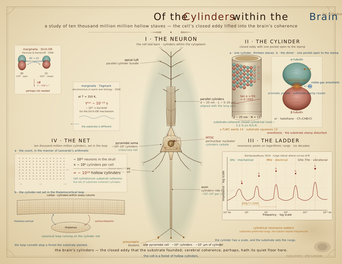
Penrose and Hameroff proposed that microtubules hold a coherence relevant to cognition, with the coherence collapsing under quantum-gravitational objective reduction. The substrate framework keeps their intuition about the object — microtubules — and replaces their mechanism entirely. The microtubule wall is a locked, closed-cylindrical modon: one of the substrate’s chirality-coherent sheets, pinned by the bridge equation’s dimensionless ratio f/\pi. The brain’s \sim 10^{16} such cylinders — roughly 10^4 per neuron — form a substrate-coherent array embedded in every neuron’s cytoplasm, an organ-scale scaffold that the cortex’s thalamocortical loops, gamma/theta rhythms, and topographic maps run on top of.
This leads to a mechanism for general anaesthesia — the most reliable experimental handle on consciousness there is — that explains, from one simple picture, a set of pharmacological facts that has genuinely puzzled the field for over a century. Follow that mechanism back to its source and it runs through the same f/\pi locking the bridge equation derives from cosmology. This chapter follows the thread from Penrose’s original intuition, through the geometry, to the anaesthesia mechanism, and back down to that equation.
Four Differences From Penrose-Hameroff
The substrate’s coherence is not quantum superposition. It is substrate-coherent structure that the cylinder’s closed geometry enforces by being substrate-locked — thermal noise can disrupt a tubulin conformation, depolymerise the cylinder, or deflect its mechanical state, but there is no superposed state for it to decohere. The framework needs no gravitational objective reduction: the substrate’s own non-Markovian dynamics, at the regime the microtubule-highways chapter works out, are already non-algorithmic in the sense Penrose’s Gödel argument gestures at, without requiring quantum-gravitational specifics to get there. The reading operates at the array scale rather than the single-cylinder scale: it is the \sim 10^{16}-cylinder population’s contribution to the brain’s organ-scale state that does the work, not any one tube’s supposed computing capacity. And the coherence is a condition maintained continuously with metabolic throughput. That property lets an anaesthetic disrupt it temporarily. The coherence is maintained, only the ability to access it is disrupted.
The last of those differences decides the shape of the two readings. Orch OR has a clock to beat: a superposition must survive long enough to reduce, and every version of the theory since 1996 has been a search for a shorter one — from the 25 ms of a gamma cycle, through the microsecond decoherence estimates of Hagan, Hameroff, and Tuszynski, to the megahertz and terahertz events of the current time-crystal picture, each step moving the reduction to a faster rung so it can fit inside the thermal budget.1 The substrate reading has no such clock, because nothing in it is waiting to collapse. The coherence is a lock that is held — re-bought continuously from the energy budget, the way a laser’s is — and the question it answers is not “how long can this survive?” but “how much loading breaks it?”, which is the question the anaesthesia section turns into a number.
The Geometry of the Microtubule
The microtubule-highways chapter works the microtubule out as a closed cylindrical modon: a hollow tube \sim 25 nm across, built from N = 13 protofilaments of α/β-tubulin heterodimers stacked head-to-tail, with the filament count itself predicted by the bridge equation.
Two well-known negative results have argued that no warm biological system could sustain the coherence this picture needs, and they disagree about which direction the failure runs: Tegmark (2000) found the brain too hot; Reimers and colleagues (2009) found it too cold. Both misses are identifiable. Tegmark computed at thermal equilibrium, which for living tissue is the condition of death — a system held far from equilibrium by a steady energy supply can condense into a coherent mode at temperatures an equilibrium calculation forbids.2 Reimers modeled a one-dimensional filament, not a closed tube — and open-helix versus closed-cylinder is exactly the distinction the microtubule-highways chapter derives: the open helix locks at \tan\alpha = f, the closed cylinder at \tan\alpha = f/\pi, with one factor of the Gauss solid angle absorbed by the cylindrical symmetry. One calculation assumed the wrong thermodynamic regime; the other assumed the wrong topology. Neither touches the object this chapter describes.
Holding \sim 10^{16} such walls in mutual coherence is a maintained condition — the same driven-dissipative regime the time-crystal and laser chapters already needed, run here on the ATP/GTP throughput the cytoskeleton already draws on. It gives a plausible structural job to a well-known number: the brain spends \sim 20\% of the body’s resting energy on \sim 2\% of its mass, mostly not on spiking but on baseline upkeep — the standing cost, on this reading, of holding the scaffold far from equilibrium.
Penrose-Hameroff, Reread
Penrose-Hameroff rests on three commitments: (1) consciousness involves a non-algorithmic process, argued from Gödel; (2) microtubules are its cellular substrate; (3) quantum-gravitational objective reduction, at the Penrose self-collapse timescale \tau \sim \hbar/E_\text{grav}, supplies the non-algorithmic dynamics. The substrate framework keeps (1) and (2) and drops (3).
and (2) survive because Hameroff’s intuition about the object was right: the microtubule is the cell’s clearest substrate-coherent organelle at the scale and abundance the argument needs, and the substrate framework supplies its own non-algorithmic dynamics without needing quantum gravity to get there — not Gödel-undecidability itself, but substrate dynamics that do not reduce to a lattice approximation at any single scale.
is dropped because nothing requires it. The Penrose timescale is a calculated quantity from a specific quantum-gravity model with no observed phenomenon that needs it, and the Tegmark calculation gives ordinary quantum-mechanical superpositions far less lifetime than Orch-OR’s mechanism requires regardless of which gravity model is used. What survives the swap are the Penrose-Hameroff empirical predictions, which stay sharp: anaesthetics should preferentially target tubulin and aromatic-pocket structures; microtubules should show multi-decade resonance structure; MT-disrupting drugs should disrupt consciousness; consciousness should track MT density. The rest of the chapter develops the cleanest of these.
The Tubulin Bit: Eighty-Six Rings and One Pocket
Hameroff’s unit of state has always been the single tubulin dimer, and his current description of it is worth taking exactly as he gives it: each dimer holds 86 π-electron resonance clouds — the aromatic rings of its phenylalanines, tyrosines, and tryptophans — which oscillate coherently and act collectively, and the dimer’s state is the orientation of the dipole that collective oscillation carries, one bit per dimer written along the lattice’s helical pathways.3 The count is exact. Human α-tubulin (TUBA1A, 451 residues) carries 20 phenylalanines, 4 tryptophans, and 19 tyrosines; human β-tubulin (TUBB, 444 residues) carries 23, 4, and 16 — forty-three rings on each monomer, eighty-six on the dimer, with a further twenty-three histidines if the imidazole is counted.
In this framework that description is already the definition of a stamp. The aromatic-pockets chapter writes any aromatic assembly as a sum over its rings, \Phi = \sum_a R_a\,\phi_{r_a}(\vec r - \vec r_a), one toroidal ring current per residue on the molecule’s own scaffold — and read off, that sum is a long vector, one coordinate per ring. A codon is a stamp of three rings on a fixed helical scaffold; the nicotinic cage is five on an evolved one; the tubulin dimer is eighty-six on a scaffold the wall geometry fixes. Its dipole — of order a thousand debye — is the leading moment of that stamp, and “dipole orientation” is the stamp’s phase relative to the counter-rotating pair the closed wall manufactures for itself. So Hameroff’s bit is, in the framework’s terms, one coordinate a_n of the wall’s long vector. A microtubule a micrometre long holds about 1{,}600 of them (thirteen protofilaments, 125 dimers each), and its state is that register. This is what the paper means by calling the microtubule the array’s scaffold rather than its computer: a register is not a processor, and 10^{16} of them is a very long vector.
What the dipole picture lacks — and what the framework adds — is a reader. A bit that flips under London forces has no input; it is a state without a signal. The framework’s reader is a ring that holds a stamp in opposite phase and computes the overlap \langle \Phi_\text{pocket} \mid \Phi_\text{occupant} \rangle against whatever sits in it, and cell biology’s own two-state tubulin is the cleanest case anywhere of an occupant setting a state. GTP-tubulin and GDP-tubulin are different conformations — the straight lattice of the growing cap and the strained lattice behind it — and the whole difference between them is one phosphate on a guanine sitting in the exchangeable site.
The structures say what that reader is built from, and the answer is not the one this chapter first assumed. An earlier draft of this section described the dimer’s pockets as lined with the dimer’s own rings, on the model of the nicotinic cage. They are not. Measured as the aromatic fraction of a pocket’s 6 Å lining divided by the aromatic fraction of the chain the pocket sits in — a scale-free statistic that asks whether selection concentrated rings at this site or merely left the local average alone — the nicotinic agonist site runs at 3.9×, seven rings crowding one small cation. Every pocket in tubulin runs below one:
| Lining | Aromatic | Enrichment | |
|---|---|---|---|
| Nicotinic agonist site (AChBP, 1UW6) — for comparison | 17 | 7 | 3.87× |
| Tubulin N-site, guanine ring (α, non-exchangeable) | 13–17 | 1 | 0.47–0.61× |
| Tubulin E-site, guanine ring (β, exchangeable) | 15–16 | 1 | 0.52–0.56× |
| Tubulin taxane site (taxol) | 36 | 2 | 0.45× |
| Tubulin colchicine site (DAMA-colchicine) | 33 | 2 | 0.50× |
Tubulin’s pockets are aromatic-poor — roughly half the ring density of the protein around them. There is no cage here, and the chapter is better off saying so.
What each nucleotide site holds instead is a single ring, and it is the same ring twice: a tyrosine stacked face-to-face on the guanine, αTyr224 and βTyr224 in the crystallographers’ numbering (βTyr222 in human TUBB). It is the only aromatic within 6 Å of either base. And the two copies are not the same stack.4
| Structure | Resolution | α site (sep., interplane) | β site (sep., interplane) |
|---|---|---|---|
| 6S8K | 1.52 Å | 3.74 Å, 12.9° | 3.97 Å, 22.8° |
| 4I4T | 1.80 Å | 3.76 Å, 12.3° | 3.93 Å, 22.1° |
| 1JFF | 3.50 Å | 3.80 Å, 6.2° | 4.09 Å, 32.7° |
The α stack is flat and tight; the β stack is tilted about ten degrees further open and sits a fifth of an ångström back. The split reproduces in three independent crystals with unrelated ligands bound elsewhere, and it survives the drop from 1.5 Å to 3.5 Å. An archaeal tubulin homolog separated from ours by something like two billion years does the same thing with a phenylalanine — 3.63 Å, 8.5° — at the structurally equivalent position, its local sequence F–S–D–L–N tracking β-tubulin’s Y–G–D–L–N. The single stacked ring is older than the microtubule.
That reads better than a cage would have. Five rings are what selection builds when the question is which ligand arrived and the answer has to separate epibatidine from choline across four orders of magnitude. One ring laid flat on one base is what it builds when identity is never in doubt and exactly one bit is at stake: the guanine is always guanine, and the only question the site ever asks is whether it carries two phosphates or three. The two copies then differ in precisely the way the framework’s two roles require. The α site is a lock closed once and never reopened — its GTP is non-exchangeable, buried at the intradimer interface behind the β subunit, and its ring reads flat. The β site is the writable coordinate, and it is held at the looser angle a reader needs if it is ever going to let the occupant go. In the lattice that site is only completed when the next dimer’s α subunit caps it, so the reader closes when the polymer closes and hydrolysis follows assembly rather than preceding it.
The signal Hameroff’s picture is missing therefore survives the correction, and the correction is to the reader’s size, not to its existence. The dimer’s state is not a dipole that happens to point one way; it is a match score, and the occupant of the pocket is the input. One ring holding one base in opposite phase is the locking pole at the smallest expression it has — the codon stamp’s base-pair closure with one half of the pair supplied by the protein instead of by a second base.
The written coordinate is not only a conformation of one dimer; it is legible on the lattice. Cryo-electron microscopy resolves two discrete lattices, an extended one with a dimer repeat of 83.2 Å in the GTP-like state and a compacted one at 81.5 Å after hydrolysis, and localises the 2 Å difference at the longitudinal interface next to β’s nucleotide, with α-tubulin’s N-terminal domain shifting and twisting toward the minus end as the E-site is read.5 One phosphate at one ring moves the whole protofilament by two ångströms per dimer. That is what a register bit with a mechanical readout looks like.
The wall then turns out to carry a second discrete variable that the chapter did not ask for. Debs and colleagues refined a thirteen-protofilament microtubule protofilament by protofilament instead of imposing helical symmetry, and found that the lateral hinge between neighbours — the M-loop of one dimer set into the H1′–S2 and H2–S3 loops of the next — does not take a continuum of angles. It takes one of two, a low-curvature and a high-curvature conformation of the M-loop about nine degrees apart, and the wall-angle distribution is bimodal at every protofilament number they measured.6 Three properties of that variable are the ones a register wants. Neighbouring hinges are strongly anticorrelated — two adjacent protofilaments counter-rotate while the rest of the wall holds still — and the correlation along a protofilament is positive and still non-zero thirty subunits away, a quarter of a micrometre. A perfectly cylindrical wall is, in their words, energetically and entropically penalised. And the seam is where the count is settled: thirteen protofilaments cannot close on one state, so the wall mixes them, and with the two measured angles the closure takes eight low and five high, the few degrees of deficit absorbed by the seam, which the same study finds buckling a further twelve degrees in a third of the population and Taxol pinning shut. Eight and five of thirteen is the split the highways chapter already found in the A-lattice’s generative helix, and the chapter records the coincidence without leaning on it: Debs reports the two peak positions but not the population fraction, and the arithmetic here is closure, not measurement. What is measurement is the pair of facts a substrate register needs — a two-state lateral contact, and a correlation length along the tube that is long compared with the dimer and short compared with the cell.
The anaesthetic enters the same mechanism — though, on the census above, not at this pocket. A xenon atom inside a ring’s reach has no ring, no lobe, no stamp to hand back; it loads the reader — polarizable enough to sit and to bend the reader’s flow into itself — and returns nothing, so the coordinate is neither written nor left alone but detuned. Hameroff’s account of anaesthesia, that the agent’s London-force interaction disperses the dipoles, is the framework’s loading term under another name, and it is right as far as it goes. What it cannot say is why a chemically inert atom is the best anaesthetic there is, why a plug that merely filled the pocket would not work, or why an odd-mass isotope of the same atom works less well. The distinction between a participant that closes the reader and a spectator that loads it is what carries those three, and the rest of the chapter runs it to the numbers. Where the volatile agents actually sit on tubulin is a separate structural question, and the enrichment statistic turns it into a sharp one: if the loading picture is right, the sites an anaesthetic occupies should come out aromatic-enriched exactly where the nucleotide and drug sites come out depleted. There is exactly one residue-level anaesthetic site on tubulin in the literature, and it points the right way. Emerson and colleagues photolabelled tadpole brain with an azido-anthracene anaesthetic, found tubulin as the target, and located the adducts near the colchicine site: two on non-aromatic residues of β’s S10 strand, and one on a tryptophan of α-tubulin’s H11′ helix, at the α–β interface behind β’s H8.7 That tryptophan does not sit alone. Within 6 Å of it in 1SA0 and 6S8K are αPhe404, αHis406, αTyr408 and βPhe262 — four rings around the one residue an anaesthetic has ever been pinned to on tubulin, about 1.6\times the chain’s aromatic fraction, against the 0.5\times of every nucleotide and drug pocket in the table above. The number is stated with its weakness: three of those rings are sequence neighbours, and without a structure of the bound anthracene this is a census of a residue’s surroundings, not of a pocket. What is not weak is the identity of the residue. αTrp407 is one of the eight tryptophans per dimer whose ultraviolet transition carries the collective response the array section rests on, so the one anaesthetic ever located on tubulin landed on an emitter of the network Kalra and colleagues saw damped.
Emerson’s own in-vitro data add a second point the chapter has to carry rather than tidy away. Isoflurane and propofol promote tubulin polymerisation, much as taxol does, while the anthracenes and the neurosteroid inhibit it, and the authors conclude that destabilising the polymer is not, by itself, the mechanism. The loading picture agrees, and says why. A spectator detunes a reader; it does not have to unbuild the wall. A stabiliser that antagonises anaesthesia — epothilone in Emerson’s tadpoles and in Khan’s rats — is holding the reader’s tuning, the lattice conformation the coordinate is written into, not the polymer’s length, and the variable the predictions below are stated in is that tuning.
The Rings on the Wall
Everything above is about one ring or one pocket. The register is the wall, so the last thing this section does is put every ring on it. The census script’s companion, scripts/tubulin-stamp/ring_atlas.py, takes the dimer as Debs and colleagues deposited it from their protofilament-refined Taxol lattice (6WVL, chains A and B), reads the lattice constants off their own coordinates rather than off a textbook — the low and high hinge screws at 23.7° and 33.4° with a lateral rise of 9.5 Å, and a dimer repeat of 83.3 Å from the two longitudinal neighbours deposited as 6WVR — and builds a thirteen-protofilament B-lattice with one seam, once at the ideal 27.7° and once with the two measured hinge states mixed eight and five, the residual closing at the seam. Each of the dimer’s rings is placed with its centroid and its normal, which in the aromatic-rings chapter is the axis of the ring’s torus. The deposited model resolves 107 of them: 76 phenyl (phenylalanine and tyrosine), 8 indole, 23 imidazole — 84 of the eighty-six once histidine is set aside. The output is one row per ring, 5{,}564 per four-dimer lattice, with its radius, azimuth and height, the orientation of its axis in the wall’s own radial–tangential–axial frame, its nearest ring, and its role. Six things the atlas says are worth recording here, and the wall simulation below is built on the same file.
Interactive: open the microtubule simulation — the thirteen-protofilament wall rebuilt in the browser from the atlas’s own dimer and lattice constants, every ring drawn as the ring simulation’s toruses on its measured centroid and normal. Fade the protein and the rings are what is left; peel the wall by radius and the two faces separate; unroll it and the seam and the five high contacts show; tryptophans only draws the one network that is not connected. The register view puts one dimer between its lateral and longitudinal neighbours: switch the E-site to GDP and the interface compacts; switch the hinge to its high state and the only ring pair across the contact barely moves. The statistics are recomputed live and checked against the atlas by &test=atlas.
| Rings | Radius | Torus axis | Where they are |
|---|---|---|---|
| αTyr224, βTyr224 | 105–106 Å | tangential | the two nucleotide stacks; β’s also at the inter-dimer interface |
| αTyr282, αHis283, βTyr283 | 90–95 Å | axial | M-loop face of the lateral hinge, lumen side |
| αPhe87, αHis88, βPhe87 | 99–101 Å | axial | H2–S3 face of the lateral hinge, lumen side |
| βHis229, βPhe272, βPhe83 | 92–102 Å | mixed | taxane site, lumen side |
| αTrp346 | 128 Å | tangential | inter-dimer interface, against the next β’s Phe404 |
| αTrp407 and its four neighbours | 122–130 Å | radial | Emerson’s anaesthetic adduct, outer surface |
| the eight tryptophans | 100–129 Å | tangential or radial, none axial | seven of eight on the outer shell |
The wall is fifty ångströms thick in rings, and the two faces have different jobs. Ring centroids run from 83 to 134 Å from the axis. The lumen face, inside 100 Å, carries both faces of the lateral hinge and the taxane site — every ring that touches the wall’s mechanics. The outer face, beyond 120 Å, carries seven of the eight tryptophans and the anaesthetic adduct — the rings that touch its optics. Patwa, Babcock and Kurian find their superradiant states weighted toward the exterior surface and their long-lived subradiant states toward the lumen; the atlas says why, since the emitters are outside.
The ring axes are isotropic. Across the dimer 36\% of ring axes point radially, 32\% tangentially, 33\% along the tube. The wall has no preferred ring orientation, which means that if it is a phased array it is phased by the stamp’s phases, not by aligned dipoles — the two toy geometries Patwa and colleagues test, every dipole axial or every dipole tangent, are both idealisations the real wall does not adopt. The stamp sum \Phi = \sum_a R_a\,\phi_{r_a} is written with a coefficient per ring for exactly this reason.
The hinge’s two states are not two ring couplings. This is the atlas’s honest negative. The only ring pair that faces across the lateral contact is αHis283 on the M-loop against αHis88 of the neighbour’s H2–S3 loop, edge-on at 6.0 Å and 58° in the low state and 5.7 Å and 56° in the high — a change of a third of an ångström and two degrees for a nine-degree hinge. The two-state variable Debs measured is a loop geometry, and the rings on both faces of the hinge sit with their axes along the tube on the lumen side. The chapter therefore does not claim the wall’s second register is a ring-current state. It claims the register exists, is discrete, and is anticorrelated between neighbours, and leaves what sets it to the loops.
The written coordinate’s ring sits at the interface that compacts. βTyr224, the E-site stack, is within 5 Å of the next dimer’s α-tubulin; so is αTrp346, one of the eight emitters, face-to-edge against the next dimer’s βPhe404 at 6.5 Å; and αTyr262 lies near-parallel, 15°, to the next dimer’s βHis406 at 7.4 Å. The 2 Å compaction the cryo-EM localises at this interface moves the reader ring, an emitter and a near-stacked pair together, which is the coupling between the pocket’s bit and the wall’s optics that the section has been assuming and can now point at. Debs’s own Taxol lattice, read off 6WVR, sits at the extended 83.3 Å repeat, consistent with their reservation that compaction is not universal.
The full ring network is connected and the tryptophan network is not. The median distance from a phenyl ring to its nearest ring is 5.3 Å, every indole has another ring within 7 Å, and three-quarters of the phenyls do. The nearest tryptophan to any tryptophan is 14–15 Å away. Patwa and colleagues keep eight rings of the hundred and find a nearest-neighbour coupling small against k_BT; the stamp keeps them all, and at van der Waals contact. This is the concrete content of the claim that the dimer is a stamp of eighty-odd rings rather than an antenna of eight.
And the two-state lattice closes. With the five high-curvature contacts spread as evenly as thirteen allows — every second or third contact, the golden spacing — twelve contacts sum to 333° and the seam takes 27°: three degrees more curved than the low state and a degree short of the symmetric 27.7°. Debs finds the seam on average three degrees more curved than symmetric, so the construction lands on the right side of the low state but short of the measurement, and the chapter records it as a construction, not a result; the full comparison needs the population fraction their figures hold and the text does not print.
Anaesthesia
Interactive: open the site view of the microtubule simulation — the anaesthetic where it has actually been found on tubulin: Emerson’s αTrp407 and its four neighbouring rings on the outer face, with a sealed xenon atom at the cluster, the rings’ flow falling into it and not returning, the five readers detuning; any noble gas at any pressure, the plug, and the odd-mass isotope, with the enrichment census beside the calibration curve. The array view runs the Kuramoto lock over every dimer’s cluster on the real lattice and loses it on the straight line \sum_i c_i/\mathrm{MAC}_i = 1, with the stabiliser acting at the taxane site on the opposite face. The earlier pocket simulation, with the spectator in the nicotinic cage, is kept below for now: open the aromatic pocket simulation — the nAChR aromatic cage of the aromatic-pockets chapter, drawn from PDB 1UW6 as the ring simulation’s toruses, with a xenon atom where the ligand was: a sealed grey sphere with no phase colour, the cage’s flow bending into it and never returning, the wraps roughening and the readers’ phases wandering. Swap in any of the noble gases at any partial pressure (the measured pressures are the inputs; the chart shows them against polarizability with helium sign-reversed), a steric plug or a rival stamp to see what the claim is not, and the odd-mass isotope to see the parity penalty. The array view runs the Kuramoto lock of a microtubule’s readers live and shows why the doses add.
In a nutshell, when an aromatic pocket - a reader built for participants - is given a spectator, coherence is temporarily disrupted. This clearly explains anaesthesia, a well studied molecular intervention that produces a reversible loss of consciousness. The Meyer-Overton correlation shows potency by lipid-water partition (1899–1901), the minimum-alveolar-concentration scale, and consistent action across vertebrates, invertebrates, and even plants. The aromatic pockets are substrate-stamp readers, sites where the substrate’s chirality-coherent structure matches a ligand’s stamp through vortex/breathing-mode coupling. This data was tested against the nAChR at \rho = +0.905 Spearman correlation across 8 ligands.
The noble gases have a sealed shell - all orbital boundaries are spectators so there is no way to make a lock inside the pocket that requires participants. They need enough diffusivity to hold the pocket open with no signal.
Three Observations Need to Fit
Wiest’s review, building on Eger and colleagues’ systematic analysis,8 lays out three facts that have sat awkwardly together for decades. Meyer-Overton: potency tracks oil/water partition across several orders of magnitude, implying a weak dispersion coupling rather than an ionic or lock-and-key bind. Species invariance: the effective dose barely varies across species, despite enormous variation in every candidate ion channel. Additivity: half an effective dose of isoflurane plus half an effective dose of cyclopropane is one effective dose — even though isoflurane strongly activates GABA-A receptors and cyclopropane barely touches them. Eger and colleagues worked through the combinations systematically and found no single channel, and no combination of channels, that reproduces the pattern.
Additivity shows that two agents with entirely different molecular binding profiles combine exactly one-for-one. This strongly suggests a single scalar downstream that drives the mechanism, and that is the aromatic pocket.

The Plug in The Pocket
The chapter on noble gases shows that heavier noble gasses increase in their soft boundary nature, increasing diffuseness. Xenon abolishes consciousness at about 70\% of an atmosphere. Noble gases have closed shells, no open lobes so that all boundaries are spectators. These form a surface already closed on itself, which can exclude but never merge. The aromatic pocket in the substrate shows recognition as a lock, one cage ring and one ligand lobe closing in opposite phase. Together the anaesthetic is clearly a spectator introduced into a pocket reader that is built for participants - the null signal.
It must be polarizable enough to sit in the pocket and load it, and sealed enough that it can never close the lock. The cage is neither blocked nor occupied by a rival — it is detuned.
The spectator view draws exactly this contrast: the same cage that closes a lock on nicotine is loaded by xenon and closes nothing, while a plug leaves it coherent and a rival stamp is answered. This is neither competitive antagonism (anaesthetic affinities are far too weak) nor steric blockade (they are small, reversible in seconds to minutes, and leave the protein’s chemistry intact). It is the channel-with-memory failure mode — a rough wrap where the structure needed a smooth one — applied simultaneously across a very large number of small readers. And it turns Meyer-Overton from a fact about lipids into a fact about polarizability per unit volume, the same coupling constant read through two instruments; the binding sites are now understood to be protein cavities rather than bulk lipid, and the correlation survived the move because dispersion into a cavity scales with polarizability just as dissolution into a lipid does.
The Noble-Gas Series Is the Calibration Curve
This data then shows the strong correlation for noble gases, their diffusivity and their behavior:
| Gas | Polarizability (ų) | Anaesthetic behavior |
|---|---|---|
| Helium | 0.205 | Non-anaesthetic; at pressure runs the effect backwards |
| Neon | 0.396 | Non-anaesthetic |
| Argon | 1.64 | Anaesthetic at \sim 39 atm |
| Krypton | 2.48 | Anaesthetic at \sim 4.5 atm |
| Xenon | 4.04 | Anaesthetic at \sim 0.95 atm (mouse); 0.6–0.7 (human) |
Five points, monotone, spanning a factor of \sim 41 in required pressure across a factor of \sim 2.5 in polarizability — and, crucially, crossing zero. Helium reverses anaesthesia under pressure and drives high-pressure nervous syndrome, which is why the helium chapter recorded it as a sign-reversed control rather than a null; a series that changes sign is far stronger evidence than one that merely trends. Xenon is the sharpest single case in all of consciousness pharmacology: an atom with no participants, no vacancies, no bonds and no chemistry of any kind reversibly abolishes consciousness and gives it back. Under a loading reading, that is the purest instance available.
Why the Doses Add
The additivity result follows from the kuramoto lock also used in the superradiance chapter. The collective-emission condition \tau_R < T_2^* is the Kuramoto lock threshold K > |\Delta\omega| — coupling must beat detuning — with 1/T_2^* the ensemble’s dephasing rate.
Give an array of readers a baseline dephasing rate 1/T_{2,0}^*, and let each anaesthetic agent i, at concentration c_i, add its own loading contribution \gamma_i c_i. Dephasing rates are rates: they add. The array holds its lock while
\frac{1}{T_{2,0}^*} + \sum_i \gamma_i c_i \;<\; \frac{1}{\tau_R},
and consciousness is lost when the sum crosses over. Writing \mathrm{MAC}_i for the concentration at which agent i alone reaches the crossing, the mixture then reaches it exactly when
\sum_i \frac{c_i}{\mathrm{MAC}_i} \;=\; 1.
The array view of the simulation computes this rather than asserting it: a microtubule’s readers in a Kuramoto lock with Lorentzian detuning, each agent adding phase noise to its own pattern of readers, and the lock lost on the straight line \sum_i c_i/\mathrm{MAC}_i = 1 — with a toggle showing the bowed curve a combination that added variances instead of rates would give, and a scaffold slider that moves MAC with the coupling. That is the additivity law as measured, and it drops out independently of every \gamma_i — independently of how each agent distributes itself across receptor types, aromatic pockets, and tubulin sites. Isoflurane’s strong GABA-A action and cyclopropane’s weak one are differences in which readers each agent roughens; they are invisible to a sum that only counts total dephasing. Heterogeneous chemistry going in, one scalar coming out — which retrodicts the fact that left Eger and colleagues perplexed rather than merely accommodating it. The same structure gives species invariance for free: the reserve is a property of the substrate-coherence architecture, common to a rat and a person alike, and \gamma_i is a property of the molecule; neither term depends on a species’ particular ion-channel inventory, so MAC has no strong reason to vary across species, and it does not.
The Chain, With a Measurement at Almost Every Link
What makes this more than a story is that Wiest’s review supplies published measurements at four of the five links.
The intruder partitions into hydrophobic pockets rich in delocalized electrons. Craddock and colleagues’ quantum-chemical modelling found several volatile anaesthetics’ potencies predicted by their binding affinity to delocalized-electron sites in tubulin — essentially reproducing Meyer-Overton from a microtubule-only assumption.9 “Delocalized electron sites” is this framework’s aromatic π-system under another name.
The loading damps the wall’s coherent energy transport. Kalra and colleagues measured electronic energy migration in microtubules directly, and found it dampened by inhalational anaesthetics10 — the single most direct measurement of the proposed mechanism that exists: the intruder goes in, and the wall’s coherent transport degrades.
The transport being damped runs on an aromatic network. The emitters are tryptophan, the most polarizable aromatic residue in the cell’s aromatic inventory — the same residue carrying the array’s collective optical response, below.
Stabilising the scaffold raises the anaesthetic requirement, and the sign is right. Wiest’s own laboratory gave rats the brain-penetrant microtubule stabiliser epothilone B and found significantly delayed loss of consciousness under isoflurane, at Cohen’s d = 1.9.11 A clinical series found slight anaesthetic resistance in surgical patients on the poorly brain-penetrant taxanes, consistent with the same mechanism at low exposure. A “microtubules are transport tracks” reading predicts nothing of the sort — stabilising the tracks should not change anaesthetic sensitivity at all — and the obvious indirect route is independently closed, since isoflurane’s inhibition of synaptic transmission is known not to run through microtubules.
Only the fifth link — array coherence quality to organ-scale integration to the presence or absence of experience — remains unmeasured, and it is the one the framework is least able to instrument.
The Isotope Result Is a Parity Experiment
Li and colleagues showed that nuclear spin reduces the anaesthetic effect. They show a roughly \sim 45\% loss of potency was possible.12
This actually comes from the change in boundary-parity of the isotopes used in the experiment, not the spin itself. A neutral atom’s fermion count is Z protons plus Z electrons plus N neutrons, so its parity is the parity of N, which is the parity of the mass number. Odd mass number, half-integer nuclear spin, and odd fermion count come together. ^{132}Xe and ^{134}Xe are even-parity bosons. ^{131}Xe and ^{129}Xe are odd-parity fermions.
From the boundary-parity rule: an even-parity object’s internal flow matches the background substrate, so it passes through the substrate transparently — no net polarization, no exclusion, while an odd-parity object’s internal flow opposes it at the outermost boundary, creating a persistent asymmetry in the local substrate.
Xenon works because it is a perfect spectator — a sealed body with no ring current, no handedness, nothing to hand the cage back (odd-mass xenon in the simulated pocket carries the parity swirl, nests less quietly, and dwells less). The effectiveness of the anaesthetic comes from the purity of that null, and boundary parity reduces the purity, the noticed reduction. An even-parity xenon is transparent to the substrate and is the cleanest anaesthetic. An odd-parity xenon has the persistent asymmetry that will nudge it along from the pocket. It is still a spectator, just a slightly worse one.
Why a sealed shell does not screen it. The hydrogen chapter sets the framework’s rule for when a nuclear property reaches the outside world — “which half you get is decided by whether there is anything between the nucleus and the world” — which is why isotope effects are a precision technique everywhere except hydrogen, and xenon has as much shell in between as anything in biology. But that rule governs mass, and mass is screened because chemistry only ever reads the boundary. Parity is not carried at a radius. It is a count taken over the whole closed object, and a sealed shell is a term in the count rather than a barrier to it. The framework has already spent this exemption once, and prominently: ³He and ⁴He sit behind the same 1s^2 shell with the same polarizability and the same absence of chemistry, differ only in a nuclear tier the periodic table cannot see, and their superfluid transitions sit three orders of magnitude apart (closed three times). The ledger “was never counting electrons in the first place — it was counting closed surfaces, and one tier down there is another one.”
Where the penalty is paid is occupancy, not loading. The pocket is not an achiral hole. It is a counter-rotating aromatic cage built out of L-amino acids, and the aromatic-pockets chapter already leans on that in reading the enantiomer puzzle in olfaction, where the GPCR seven-TM bundle is “unavoidably chiral, the same right-handed bias that organizes B-DNA’s pitch running through every aromatic side chain in the cage.” A polarization the substrate carries has a handedness; a handed boundary either accommodates it or shears against it. So the odd-parity atom’s cost is not that it loads less — it loads identically — but that it does not nest as quietly, and the lithium chapter’s reading of transport applies unchanged one scale down: the wrap is the mover, and how the wrap is held decides how long the thing stays. A slightly rougher sit is a shorter dwell, a lower time-averaged occupancy at the same partial pressure, and a higher dose to reach the same total dephasing. In the additivity ledger above, the parity term multiplies \gamma_i through occupancy while leaving every other term where it was — which is why the isotope effect is a 45\% correction rather than the factor of 41 the polarizability axis commands across the same column.
This is no longer unsupported. When this section was first written it recorded a lone conjecture with the right sign and nothing behind it. Wang and Ozturk have since proposed, from a completely different direction, that the Li result is the nuclear-spin-dependent permeability of isotopes through homochiral media, modulating occupancy at a receptor through a Hill–Langmuir term — a spin-selective barrier at a chiral interface, with the interface read as the mouth of the pocket and the compartment behind it as the active site. Their route is chirality-induced spin selectivity, an experimentally established room-temperature effect in which chiral molecules act as spin filters. That is the same claim this section makes, in a different vocabulary, reached without any of this framework’s machinery. It also arrives with the same two negative results: the effect is not in the atom’s volume or polarizability, and it does not require long-range coherence. The radical-pair account remains live but is not the settled reading it was — it needs a radical pair inside a hydrophobic pocket with no photon to make one, and a T_2 that survives body temperature, and neither has been demonstrated.
Two predictions separate the readings, and group 18 offers exactly three tests. All three stable argon isotopes are even-mass and spin-zero, so argon cannot be tested at all; neon’s odd isotope is not anaesthetic. What remains is ^3He, ^{83}Kr, and the xenon pair already run.
Parity predicts a step; hyperfine coupling predicts a gradient. If the penalty is a parity class, every odd-mass isotope pays the same fractional cost regardless of its magnetic moment. If it runs through hyperfine coupling, the cost tracks |\mu| and the coupling’s own dependence on polarizability. The existing data cannot separate them: ^{129}Xe carries 12\% more magnetic moment than ^{131}Xe and pays 6 \pm 9 more points of ED_{50}, which is consistent with either. Higher-precision replication is the cheap test.
Krypton-83 is the sharp one, and nobody has run it. It is the only stable odd-mass krypton, at I = 9/2 and |\mu| = 0.971\,\mu_N — a larger moment than either xenon isotope, sitting on a markedly tighter boundary. Parity predicts ^{83}Kr pays xenon’s fractional penalty, moving krypton from \sim 4.5 atm to \sim 6.5. Hyperfine coupling predicts a smaller fractional penalty than xenon’s, because the coupling needs the polarizability krypton does not have. Falsified by a ^{83}Kr penalty that scales with the nuclear moment rather than holding the step, and falsified outright by no isotope effect in krypton at all.
A third test closes a loop the book already opened. ³He is odd-parity, and helium is the series’ sign-reversed control — too tight to sedate, so at depth its excitation shows through as high-pressure nervous syndrome. The same isotope pair the helium chapter used to move the superfluid transition by 10^3 should therefore also move the HPNS threshold, in whichever direction the reversed sign dictates. It is an expensive experiment and has not been done.
What is still owed is the number. The framework gets the sign, the selectivity rule, and the prediction that the effect vanishes in achiral media — but it does not derive why the penalty is 45\% rather than 5\%, because it has no coupling constant between a parity asymmetry and a chiral cage’s shear. And the whole structure rests on a single unreplicated study of eighty mice. Wang and Ozturk say the same of their own model, that everything “rests critically on the validity and reproducibility of the underlying observation,” and it is worth repeating here. What has changed is the standing of the result: it is no longer an anomaly the loading picture has no business producing. It is the loading picture’s purity claim, tested in the one way that holds polarizability exactly fixed, and passing.
The Two-Term Model
Potency, on this reading, splits into an achiral loading term — set by polarizability, the whole story for the noble gases — and a smaller stamp term that determines which readers a structured agent preferentially loads. Mirror-image molecules share identical polarizability, so the loading term cancels exactly in an R/S comparison, leaving the stamp term isolated. That resolves a real tension in the literature: enantiomeric potency differences for inhaled anaesthetics are real but modest, which pure lipid theory (predicting zero) and pure lock-and-key theory (predicting a large ratio) both get wrong, and a large achiral term plus a small chiral one gets right.
The Array That Closes the Loop
A cortical pyramidal neuron holds \sim 10^5 MT cylinders; the brain’s \sim 10^{11} neurons hold \sim 10^{16} of them altogether, from the dendritic spine (\sim 10^1) through the soma’s radiating bundle (\sim 10^4–10^5) to the axon (\sim 10^4–10^6 per caliber). Whether that count is doing any work, rather than just bookkeeping, now has a direct answer. Babcock and colleagues analysed collective ultraviolet excitation of tryptophan networks across a nested hierarchy — a single microtubule, a centriole of 27, a neuronal bundle of 91 in hexagonal packing — and found strongly superradiant states whose fluorescence quantum yield grows with network size across all three scales, at room temperature.13
That is the distinction between an array claim and a decorative one: coherence that saturated at a single cylinder would make the \sim 10^{16} count mere bookkeeping, and it does not saturate with tube count. The same group’s later calculation is specific about what does saturate: a single microtubule’s peak collective decay rate stops growing once the tube is about three ultraviolet wavelengths long, roughly 0.8\;\mum, while the quantum yield keeps rising out to twenty thousand tryptophans and the enhancement keeps rising with the number of tubes in the bundle.14 The array claim is stated in that variable. The framework already had the mathematics for it. The superradiance chapter reads collective emission as wake overlap, with locked boundaries adding as amplitudes so radiated power runs as N^2, and its central reading of the superradiant laser is that phase is safest when stored in a macroscopically occupied material mode and only transiently expressed as light. That sentence was written about strontium atoms in a bad cavity. Unmodified, it is also the claim this chapter makes about the cylinder array: the coherence lives in the array’s lock, and the UV superradiance merely reports it.
It also closes the anaesthesia argument into a loop, rather than leaving it as a list of separate facts. The emitters carrying the collective response are tryptophan; the hydrophobic pockets a volatile anaesthetic partitions into sit inside that same aromatic network; and what Kalra and colleagues measured, when they put anaesthetics on microtubules, was the damping of exactly this coherent transport. The intruder, the network it loads, and the collective response that degrades are one object described three times — and the object, all the way down, is the same closed-cylindrical geometry the bridge equation’s f/\pi locks in the first place.
The Fan and the Flip: Where the Match Is Computed
The microtubules in the brain are not laid out uniformly, and the way they are laid out is the chapter’s best clue to what the array is for. Hameroff’s slides draw the arrangement in a pyramidal neuron — the layer-5 cell type, not a shape, though the shape happens to be right: an apical tuft fanning inward through one trunk to the soma, a skirt of basal dendrites, one axon out. Nearly four decades of tracking microtubule polarity in that cell give a clean split.15 In the axon every microtubule points the same way, plus end outward, long, continuous, tau-bound: a highway. In the dendrites and soma they are short, interrupted, MAP2-cross-linked, and of mixed polarity — roughly half plus-end-out and half minus-end-out, so that a section through a dendrite finds tubes pointing both ways side by side. This is the “every other one flipped” of Hameroff’s picture, and the flip is organised: the minus-end-out population is the stable, acetylated one, the plus-end-out population the dynamic, tyrosinated one, and they sort into bundles of opposite orientation, which is how kinesin-1, preferring acetylated tubes, is steered out of dendrites and into the axon.16
The framework reads the two compartments as the two halves of one operation. The axon is the lock pole as a conduit: one handedness, one direction, a result carried away. The dendrite’s antiparallel pairs are a modon at the bundle rung — the counter-rotating pair the highways chapter found inside a single tube’s wall, here built between two tubes and held at spacing by MAP2. And a counter-rotating pair is what the coherence match is made of. The overlap \langle a \mid b\rangle = \sum_n a_n^{*} b_n has two parts: a product of one register against the conjugate of another — one vector run against the other in reverse, the “opposites attract” closure the codon stamp reads at a base pair — and a sum over all of them. An antiparallel pair of registers, bridged along their length, is the product term laid out in tubulin: one tube’s coordinates read against a neighbour’s running the other way. The fan is the sum: 10^4–10^5 synapses on some ten millimetres of branch, every branch converging on one soma. The dendritic arbor of a pyramidal neuron is the inner product drawn in anatomy — the antiparallel bundle as the multiply, the tuft as the add — and the axon carries the result. This is the cytoskeletal rung of the operation the cortical-resonator ODE runs at the column rung: the fan is a reduction, the flip is the match, and a differential-equation solver built out of coherence matches would look like this.
The cell biology of the pyramidal neuron already says the apical fan is where a match is computed. Larkum’s cellular mechanism for cortical association has the layer-5 cell receive top-down context on its apical tuft and bottom-up drive on its basal dendrites, and burst only when the two coincide within a window — a coincidence detector between prediction and measurement, which is what the prediction-engine chapter reads the canonical loop as at the circuit level.17 The framework puts the match one level down, on the mixed-polarity scaffold the apical compartment is built on.
And this is where anaesthesia lands, measured. Suzuki and Larkum drove the distal apical dendrites of layer-5 pyramidal neurons optogenetically: awake, the drive spikes the soma; under any of three chemically unrelated anaesthetics it does not — the apical tuft is decoupled from the cell body while the cell body itself still fires — and the same decoupling is produced by blocking the metabotropic receptors on the tuft or by silencing the higher-order thalamus that feeds it.18 Bharioke and colleagues found the complementary signature across cortex: under different anaesthetics, layer-5 pyramidal neurons — and no other cortical cell type — fall into global synchrony, with the change in synchrony tracking the loss and recovery of consciousness.19 Neither result was obtained with the cytoskeleton in mind, and neither derives the loading picture. But together they identify the compartment: the link an anaesthetic cuts is the apical fan’s coupling into the soma — the sum failing to reach the result — in exactly the cell whose apical compartment is built on the mixed-polarity, MAP2-bridged register arrays. The one-cell form of this chapter’s array claim is that the tuft is the reader and the anaesthetic silences the read.
What the Substrate Adds to Penrose’s Vision — and What It Still Can’t
Penrose’s original argument wanted more than a mechanism for coherence: it wanted an objectively real whole for a unified conscious moment to correspond to, because ordinary classical physics has none. A classical system reduces completely to local interactions among neighbours, so any larger-scale object — a brain, a tornado — is eliminable from the description without loss, and an eliminable whole has no causal power. Quantum mechanics escapes that trap because entanglement supplies a holism that Bell proved no local account can reproduce.
The substrate framework escapes it too, by a different route, and has done so since the Bell chapter: the framework violates locality below the modons that constitute emergent physics, through a channel whose topological charge cannot be unwound by local fluctuations. A winding number is not a function of any local neighbourhood — reduce a modon to its constituent lattice cells and the circulation appears nowhere in the reduction, not because it is hard to compute but because it is a different kind of quantity. That makes a modon exactly the kind of object Penrose’s argument says only quantum mechanics can supply — an objective, causally efficacious whole — without superposition, without a decoherence budget, and without gravitational collapse.
What this does not buy is an explanation of experience itself. It says what the objective whole is, with a geometry and an energy budget attached; it does not say why there is something it is like to be one. That limit is stated honestly here rather than argued around.
Six Predictions
Anaesthetic potency splits into a loading term and a stamp term, and enantiomers isolate the second. The noble-gas series is the loading term’s calibration curve, already measured. The decisive test is enantiomer pairs: measured R/S potency ratios should be larger for structured intravenous agents (ketamine, etomidate) than for small halogenated inhalational agents (isoflurane), tracking how much of each agent’s potency the stamp term carries. Falsified if the stamp term adds nothing over polarizability, or enantiomer pairs show no systematic difference.
Stabilising and depolymerising the scaffold move MAC in opposite, dose-dependent directions, and the variable is the lattice conformation, not the polymer mass. Epothilone B already delays loss of consciousness at large effect size (Khan et al. 2024, d = 1.9), and epothilone D halves the anthracene’s potency in tadpoles (Emerson et al. 2013); the scaffold reading predicts this is monotone in stabiliser dose and achieved MT density, and predicts the opposite sign for depolymerisers — nocodazole, colchicine, vinca alkaloids — at sub-cytotoxic doses. It does not predict that the anaesthetics themselves depolymerise: isoflurane and propofol are mild stabilisers in vitro, and the reading needs them to be spectators in a reader, not solvents of the wall. Falsified by a single-sign, dose-independent result, by a depolymeriser that also raises MAC, or by a stabiliser that raises MAC only in proportion to polymer mass rather than to the fraction of the lattice held in the compacted, hydrolysed conformation.
Collective response scales with array size, and anaesthetic sensitivity scales inversely with it. Babcock’s enhancement (single microtubule → centriole → bundle) should continue growing with bundle size and ordered packing, and should degrade when the packing is disordered at fixed tube count; cell types and cortical regions with denser, better-ordered bundles should be correspondingly harder to push below threshold. A single tube’s enhancement is already known to saturate in length at a few ultraviolet wavelengths, and that is not the falsifier. Falsified if collective enhancement saturates with tube count at fixed packing, or is indifferent to regional anaesthetic sensitivity.
The parity penalty is a step, and krypton-83 is where it can be caught. The xenon isotope result above reads as a boundary-parity effect on pocket occupancy rather than a loading effect, so it should apply as a fixed fractional cost to every odd-mass isotope, independent of nuclear magnetic moment. ^{83}Kr, the only stable odd-mass krypton, carries a larger moment than either xenon isotope on a markedly tighter boundary, and should move krypton from \sim 4.5 atm to \sim 6.5. Falsified by a krypton penalty that scales with |\mu| instead of holding the step, or by no krypton isotope effect at all.
The dendritic scaffold, not the axonal one, sets anaesthetic sensitivity. The match is computed on the mixed-polarity, MAP2-bridged arrays of the apical compartment, so interventions that disrupt dendritic cross-linking — MAP2 loss or hyperphosphorylation, dendrite-selective depolymerisation — should lower the anaesthetic requirement, while equivalent disruption of the axonal array through tau should not; and the apical decoupling Suzuki and Larkum measured should be delayed by epothilone B in step with the loss of righting. Falsified if MAC tracks axonal integrity as strongly as dendritic, or if the stabiliser moves the behavioural endpoint without moving the apical decoupling.
Where a microtubule is a register its lattice is Fibonacci; where it is a highway it is B. The highways chapter works the arithmetic: the A-lattice’s generative helix steps five protofilaments per dimer, 138.5°, the Fibonacci convergent of the golden angle — the ladder’s anti-lock packing — while the in-vivo B-lattice is not phyllotactic and needs a seam. The sign rule predicts A-lattice content enriched in dendritic and somatic arrays and depleted in the axon. Falsified by lattice type indifferent to compartment.
The Resonance Cascade: Clocks Within Clocks
The frequency ladder Hameroff now puts at the centre of the picture comes from one laboratory. Bandyopadhyay’s group at NIMS measured alternating-current transmission across single tubulin dimers, single microtubules, and the initial segment of a neuron with four-probe nanoelectrodes, and reported the same “triplet of triplets” of resonance bands recurring at each object, shifted by three decades each time.20 Laid out with Hameroff’s assignment of which subsystem rings at which band:21
| Object | Bands reported | Hameroff’s assignment |
|---|---|---|
| Neuron, EEG | Hz · kHz · MHz | membrane and network rhythms |
| Microtubule bundle | 10^2–10^3 Hz | — |
| Microtubule | kHz · MHz · GHz | kHz: tubulin C-termini; MHz: lattice phonons and polaritons; GHz: ordered water in the hollow core |
| Tubulin | MHz · GHz · THz | THz: aromatic π-electron transitions |
Each object spans three bands, and each step up in object drops the whole triplet by a thousand. Hameroff’s phrase for it is fractal time crystal — “clocks within clocks,” the same dynamics nested at every rung — and the group also reports the megahertz triplet from the human scalp, suppressed by general anaesthetics. The measurements are unreplicated outside that laboratory and contested within the field; the chapter leans no weight on their numbers. But the shape of the claim is one the framework has a definite reading of, and the reading fixes what would count as a test.
Within one object the peaks should be harmonic; the ladder is between objects. The substrate ladder is a comb of ratios because the medium between its cutoffs has no length; a structure that has a length — a ring, a tube, a cell — rings at integers of a fundamental, which is how the highways chapter reads the count thirteen and the aromatic-rings chapter reads Hückel’s modes. A microtubule is a bounded cavity. Its kilohertz-to-megahertz peaks should therefore be the overtone and band structure of a periodic object of finite length, not rungs of a \sqrt2 comb — and that is how the group’s own collaborators fit them: Sanchez-Castro and colleagues model the microtubule as a one-dimensional crystal with an 8 nm period and reproduce the measured 10 kHz–250 MHz transmission spectrum as Bloch bands with defect cavities.22 An earlier draft of this section predicted that the peaks would cluster on the \sqrt2 comb; that was the wrong template for a cavity, and the prediction is withdrawn. What the framework does claim is the self-similarity — the same triplet recurring as the object grows — because discrete scale invariance is the symmetry the ladder is built from, and “fractal” and “scale free” are that symmetry named by the people who measured it. The ratio between rungs of this cascade is chemistry’s choice of lengths (a dimer, a tube, a cell), not the substrate’s pairing factor, so the framework does not predict the thousand.
One rung of the cascade is not set by a length. Every band below terahertz is fixed by something with a size: a C-terminal tail, a lattice period, a water column, a cell. The substrate itself owns exactly one absolute frequency, and it is the anti-phase breath of its own quantum — the dc1 Compton frequency, m_1 c^2/h = 2.03\;\text{meV}/h \approx 0.49 THz, with the lattice’s infrared floor hc/\xi = 2\pi m_1 c^2 \approx 13 meV at 3.1 THz above it. Both sit in the 10^{11}–10^{12} Hz band at the top of Hameroff’s ladder, the band he assigns to the π-electron oscillations of the eighty-six rings — the one place in the cascade where a substrate clock rather than a molecular length could set the number. That is a coincidence worth recording and not yet worth more. The terahertz band is crowded: thermal energy at body temperature is k_BT/h \approx 6.5 THz, collective protein modes fill the decade below it, and the universal low-frequency excess every protein and glass shows — the boson peak — sits near a terahertz. A substrate clock would be distinguishable from all of these by one property: it does not scale. A length-set mode moves when the protein changes; a feature at 2 meV (16 cm^{-1}) that is the same in tubulin, in a receptor cage, and in any aromatic-rich protein of any size would be the breath showing through, and one that drifts with molecular size is not. The chapter does not claim the breath is what the group measured. It notes that if the top of the cascade is real, the substrate has a number waiting there, and that the number is falsifiable by a spectrum. The breath is expressed through many layers of chemistry before it reaches an electrode, and pinning it to a peak is harder than naming the band.
The scalp signal, if real, inherits the anaesthesia ledger. The claim that megahertz triplets are detectable from the human scalp and suppressed by anaesthetics is the one piece of the cascade that touches this chapter’s mechanism directly. If it holds, the signal is a readout of the array lock, and it must then obey the lock’s arithmetic: its suppression by a mixture of agents should follow the same straight line \sum_i c_i/\mathrm{MAC}_i = 1 the additivity section derives, and its loss should be delayed by the microtubule stabiliser epothilone B by the margin Khan and colleagues measured for the righting reflex. A megahertz scalp signal that tracked a different dose law than consciousness, or was unmoved by a stabiliser that moves MAC, would be an artifact or a different object. The reported cross-cellular reach of the resonance state — spanning neurons and steering membrane voltage — is the reach the array reading needs, and the same test applies to it.

Putting the Section in Context
The thread runs from one end of the figure above to the other. Penrose wanted a physical object able to host a non-algorithmic, objectively unified process, and reached for quantum-gravitational collapse to get it. The substrate supplies that object without the collapse: a closed-cylindrical modon, locked by the same f/\pi ratio the bridge equation derives from cosmology, its non-locally-reducible winding number doing the work Penrose needed entanglement for. Follow that geometry into the brain’s \sim 10^{16}-cylinder array and it predicts, from one picture — a sealed, polarizable body loading an aromatic reader it can never lock — a set of pharmacological facts (Meyer-Overton, species invariance, additivity, the noble-gas sign reversal) that a century of ion-channel pharmacology never unified. Follow the mechanism further and it makes contact with data at nearly every link: coherent transport measured and measurably damped by anaesthetics (Kalra 2023), a stabiliser that delays unconsciousness in the predicted direction (Khan 2024), and a collective optical response that grows with array size across three architectural scales (Babcock 2024) — the same aromatic network, read three different ways, all pointing at the same object. Two pieces of Hameroff’s own recent picture then fall into place on the same reading without being asked for: the eighty-six rings per dimer are a stamp, and the pyramidal neuron’s mixed-polarity dendritic arrays against its uniform axon are the match and the result, laid out in exactly the compartment where the anaesthetic is measured to cut (Suzuki and Larkum 2020). The one result that looked like an anomaly, the xenon isotope effect, turns out to be that same purity claim tested at the only tier which holds polarizability fixed, and it holds.
That is the shape of the argument this chapter makes: not a story assembled to fit anaesthesia, but a geometry derived for other reasons — the microtubule wall, the aromatic pocket, the bridge equation’s own dimensionless ratio — that turns out to predict it. The neuron, synapse, and cortical-maps-and-rhythms chapters built the brain’s organ-scale architecture on top of this scaffold; this chapter takes it down to the smallest cellular rung that architecture rests on. What remains speculative is the top of the stack, honestly: the step from array coherence quality to the presence or absence of experience is unmeasured, and naming the topological object consciousness might correspond to is not the same as explaining why there is something it is like to be one.
The framework’s broader speculative arc continues in Lifetime of the Stamp (the codon-stamp ring-down time the framework’s biological propagation claims stand or fall on) and The Turing-Complete Cell (what long stamp lifetimes would mean for protein folding and substrate-information processing in cells).
Footnotes
Hagan, S., Hameroff, S.R. & Tuszynski, J.A., “Quantum computation in brain microtubules: decoherence and biological feasibility,” Physical Review E 65, 061901, 2002.↩︎
Fröhlich, H., “Long range coherence in biological systems,” Rivista del Nuovo Cimento 7, 399–418, 1977; Wu, T. & Austin, S.J., “Fröhlich’s model of Bose condensation in biological systems,” Journal of Biological Physics 9, 97–107, 1981.↩︎
Hameroff, S. & Penrose, R., “Consciousness in the universe: a review of the ‘Orch OR’ theory,” Physics of Life Reviews 11, 39–78, 2014; Hameroff, S., Bandyopadhyay, A. & Lauretta, D.S., “Microtubules are ‘Fractal Time Crystals’: implications for life and consciousness,” Journal of Consciousness Studies 33, 211–247, 2026.↩︎
Measured from PDB 6S8K (La Sala, G., Olieric, N., Sharma, A. et al., “Structure, thermodynamics, and kinetics of plinabulin binding to two tubulin isotypes,” Chem 5, 2969–2986, 2019); 4I4T (Prota, A.E., Bargsten, K., Zurwerra, D. et al., “Molecular mechanism of action of microtubule-stabilizing anticancer agents,” Science 339, 587–590, 2013); 1JFF (Löwe, J., Li, H., Downing, K.H. & Nogales, E., “Refined structure of αβ-tubulin at 3.5 Å resolution,” Journal of Molecular Biology 313, 1045–1057, 2001); 1SA0 (Ravelli, R.B.G., Gigant, B., Curmi, P.A. et al., “Insight into tubulin regulation from a complex with colchicine and a stathmin-like domain,” Nature 428, 198–202, 2004); 7EVC (Akıl, C., Ali, S., Tran, L.T. et al., “Structure and dynamics of Odinarchaeota tubulin and the implications for eukaryotic microtubule evolution,” Science Advances 8, eabm2225, 2022). Separation is between ring centroid and guanine-plane centroid; the angle is between the two best-fit planes. The census script is
scripts/tubulin-stamp/pocket_census.py.↩︎Zhang, R. & Nogales, E., “A new protocol to accurately determine microtubule lattice seam location,” Journal of Structural Biology 192, 245–254, 2015; Manka, S.W. & Moores, C.A., “Microtubule structure by cryo-EM: snapshots of dynamic instability,” Essays in Biochemistry 62, 737–751, 2018; Kellogg, E.H., Hejab, N.M.A., Howes, S. et al., “Insights into the distinct mechanisms of action of taxane and non-taxane microtubule stabilizers from cryo-EM structures,” Journal of Molecular Biology 429, 633–646, 2017. The compaction is one laboratory’s measurement chain, is not seen in yeast tubulin, and Debs and colleagues (below) decline to call it universal.↩︎
Debs, G.E., Cha, M., Liu, X., Huehn, A.R. & Sindelar, C.V., “Dynamic and asymmetric fluctuations in the microtubule wall captured by high-resolution cryoelectron microscopy,” Proceedings of the National Academy of Sciences 117, 16976–16984, 2020. Their deposited models of the two states, 6WVL and 6WVM, give lateral screw angles of 23.7° and 33.4° between neighbouring dimers when superposed directly, with a lateral rise of 9.5 Å, the 3-start step; the fitted peak values in the paper are 24.3° and 33.1°.↩︎
Emerson, D.J., Weiser, B.P., Psonis, J. et al., “Direct modulation of microtubule stability contributes to anthracene general anesthesia,” Journal of the American Chemical Society 135, 5389–5398, 2013. The labelled tryptophan is numbered 406 in their isoform table and is αTrp407 in the PDB numbering used here.↩︎
Eger, E.I. 2nd, Raines, D.E., Shafer, S.L. et al., “Is a new paradigm needed to explain how inhaled anesthetics produce immobility?” Anesthesia & Analgesia 107, 832–848, 2008.↩︎
Craddock, T.J.A., Kurian, P., Preto, J. et al., “Anesthetic alterations of collective terahertz oscillations in tubulin correlate with clinical potency,” Scientific Reports 7, 9877, 2017.↩︎
Kalra, A.P., Benny, A., Travis, S.M. et al., “Electronic energy migration in microtubules,” ACS Central Science 9, 352–361, 2023.↩︎
Khan, S., Huang, Y., Timuçin, D. et al., “Microtubule-stabilizer epothilone B delays anesthetic-induced unconsciousness in rats,” eNeuro 11, 2024.↩︎
Li, N., Lu, D., Yang, L. et al., “Nuclear spin attenuates the anesthetic potency of xenon isotopes in mice: implications for the mechanisms of anesthesia and consciousness,” Anesthesiology 129, 271–277, 2018. Adjacent masses carry the contrast — ^{131}Xe against ^{132}Xe differ by one neutron and by 29 points of ED_{50} — so ordinary mass-dependent kinetics are excluded by the design.↩︎
Babcock, N.S., Montes-Cabrera, G., Oberhofer, K.E., Chergui, M., Celardo, G.L. & Kurian, P., “Ultraviolet Superradiance from Mega-Networks of Tryptophan in Biological Architectures,” Journal of Physical Chemistry B 128, 4035–4046, 2024.↩︎
Patwa, H., Babcock, N.S. & Kurian, P., “Quantum-enhanced photoprotection in neuroprotein architectures emerges from collective light-matter interactions,” Frontiers in Physics 12, 1387271, 2024. Their toy model that comes closest to this chapter’s geometry — dipoles lying tangent to rings of 104 stacked at 8 nm — is the one whose yield rises with packing density. The authors state that the yield increase “does not have any classical interpretation,” because a single-photon excitation of a genuinely quantum transition is involved; the framework does not dispute that the tryptophan transition is quantum, and reads the delocalised single excitation as the probe. What holds the phase between rings across a micrometre is the question their Hamiltonian assumes an answer to, and it is the question this chapter’s lock is an answer to.↩︎
Baas, P.W., Deitch, J.S., Black, M.M. & Banker, G.A., “Polarity orientation of microtubules in hippocampal neurons: uniformity in the axon and nonuniformity in the dendrite,” Proceedings of the National Academy of Sciences 85, 8335–8339, 1988.↩︎
Tas, R.P., Chazeau, A., Cloin, B.M.C., Lambers, M.L.A., Hoogenraad, C.C. & Kapitein, L.C., “Differentiation between oppositely oriented microtubules controls polarized neuronal transport,” Neuron 96, 1264–1271, 2017.↩︎
Larkum, M., “A cellular mechanism for cortical associations: an organizing principle for the cerebral cortex,” Trends in Neurosciences 36, 141–151, 2013.↩︎
Suzuki, M. & Larkum, M.E., “General anesthesia decouples cortical pyramidal neurons,” Cell 180, 666–676, 2020.↩︎
Bharioke, A., Munz, M., Brignall, A. et al., “General anesthesia globally synchronizes activity selectively in layer 5 cortical pyramidal neurons,” Neuron 110, 2024–2040, 2022.↩︎
Sahu, S., Ghosh, S., Ghosh, B., Aswani, K., Hirata, K., Fujita, D. & Bandyopadhyay, A., “Atomic water channel controlling remarkable properties of a single brain microtubule: correlating single protein to its supramolecular assembly,” Biosensors and Bioelectronics 47, 141–148, 2013; Saxena, K., Singh, P., Sahoo, P. et al., “Fractal, scale free electromagnetic resonance of a single brain extracted microtubule nanowire, a single tubulin protein and a single neuron,” Fractal and Fractional 4, 11, 2020.↩︎
Hameroff, S., Bandyopadhyay, A. & Lauretta, D.S., “Microtubules are ‘Fractal Time Crystals’: implications for life and consciousness,” Journal of Consciousness Studies 33, 211–247, 2026.↩︎
Sanchez-Castro, N., Palomino-Ovando, M.A., Singh, P. et al., “Microtubules as one-dimensional crystals: is crystal-like structure the key to the information processing of living systems?” Crystals 11, 318, 2021.↩︎